
Figure 7:
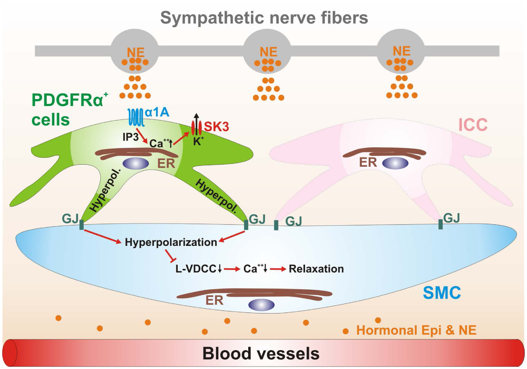
Schematic diagram depicting a novel signal pathway of sympathetic neural regulation of murine colon. Sympathetic nerve fibers, a blood vessel and the cells of the SIP syncytium (SMCs, ICC and PDGFRα+ cells) are displayed. Gap junctions (GJ) are shown between SMCs and ICC and PDGFRα+ cells in dark green. PDGFRα+ cells (pale green cell) express α1A ARs. Neuronal or hormonal norepinephrine (NE) (and possibly epinephrine; Epi) (orange circles) bind to α1A ARs, enhance Ca2+ release from endoplasmic reticulum (ER) via generation of inositol triphosphate (IP3) and activate small conductance Ca2+ -activated K+ channels type 3 (SK3; expressed robustly in PDGFRα+ cells; Fig. 1B). SK3 channels generate outward currents and induce hyperpolarization (Hyperpol.) in PDGFRα+ cells, which conducts via GJ to SMCs (pale blue cell) and probably ICC (pink cell). Hyperpolarization of SMCs reduces the open probability of L-type Ca2+ channels (L-VDCC) and decreases intracellular [Ca2+], leading to inhibition of contractions. ICC (pale pink cell) are not involved in this novel signal pathway.