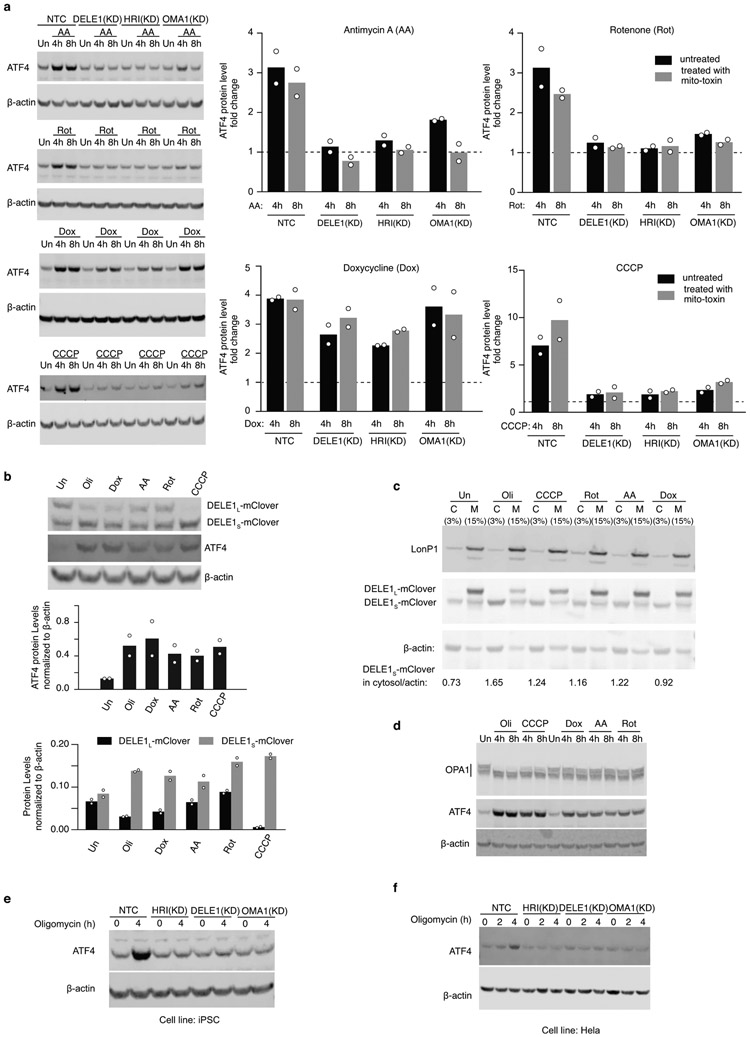
Extended Data Figure 6. Examination of mitochondrial stress response with a broad range of mitochondrial toxins and in non-HEK293T cells.
(a) Immunoblot of ATF4 in wild type, DELE1(KD), HRI (KD) and OMA1(KD) HEK293T cell lines under different mitochondrial stress conditions. Cells were left untreated (Un) or treated with 40nM Antimycin A (AA), 40nM Rotenone (Rot), 50 ug/mL Doxycycline (Dox), or 5 μM CCCP for 2 and 4 hrs. Left, representative blots. Right, ATF4 levels were quantified and normalized to β-actin (mean ± s.d., n = 2 blots).
(b) A broad range of mitochondrial toxins stimulates the accumulation of DELE1S. Cells stably expressing DELE1-mClover were untreated or treated with a panel of mitochondrial toxins for 16 h (see Methods for details) and subjected to Western blotting with antibodies detecting DELE1-mClover, ATF4 and actin. Top, representative blot. Middle, bottom, ATF4, DELE1L-mClover and DELE1S-mClover levels were quantified (mean ± s.d., n = 2 blots).
(c) Subcellular localization of DELE1L and DELE1S under a broad range of mitochondrial toxins.
Biochemical fractionation of cells stably expressing DELE1-mClover that were either treated with different mitochondrial toxins as indicated for 16 h or left untreated. β-actin and LonP1 were probed as markers for cytosol and mitochondria, respectively. Similar results obtained in n = 2 independent experiments.
(d) Examination of OPA1 cleavage under a broad range of mitochondrial toxins. Similar results obtained in n = 2 independent experiments.
(e) Immunoblot of ATF4 in wild type, DELE1(KD), HRI (KD) and OMA1(KD) in the WTC11 human iPSC line. Cells were left untreated or treated with 1.25 ng/mL oligomycin for 4 hrs. Similar results obtained in n = 2 independent experiments.
(f) Immunoblot of ATF4 in wild type, DELE1(KD), HRI (KD) and OMA1(KD) in the human Hela cell line. Cells were left untreated or treated with 1.25 ng/mL oligomycin for 2 and 4hrs.
Similar results obtained in n = 2 technical replicates.
For gel source data, see Supplementary Figure 1.

