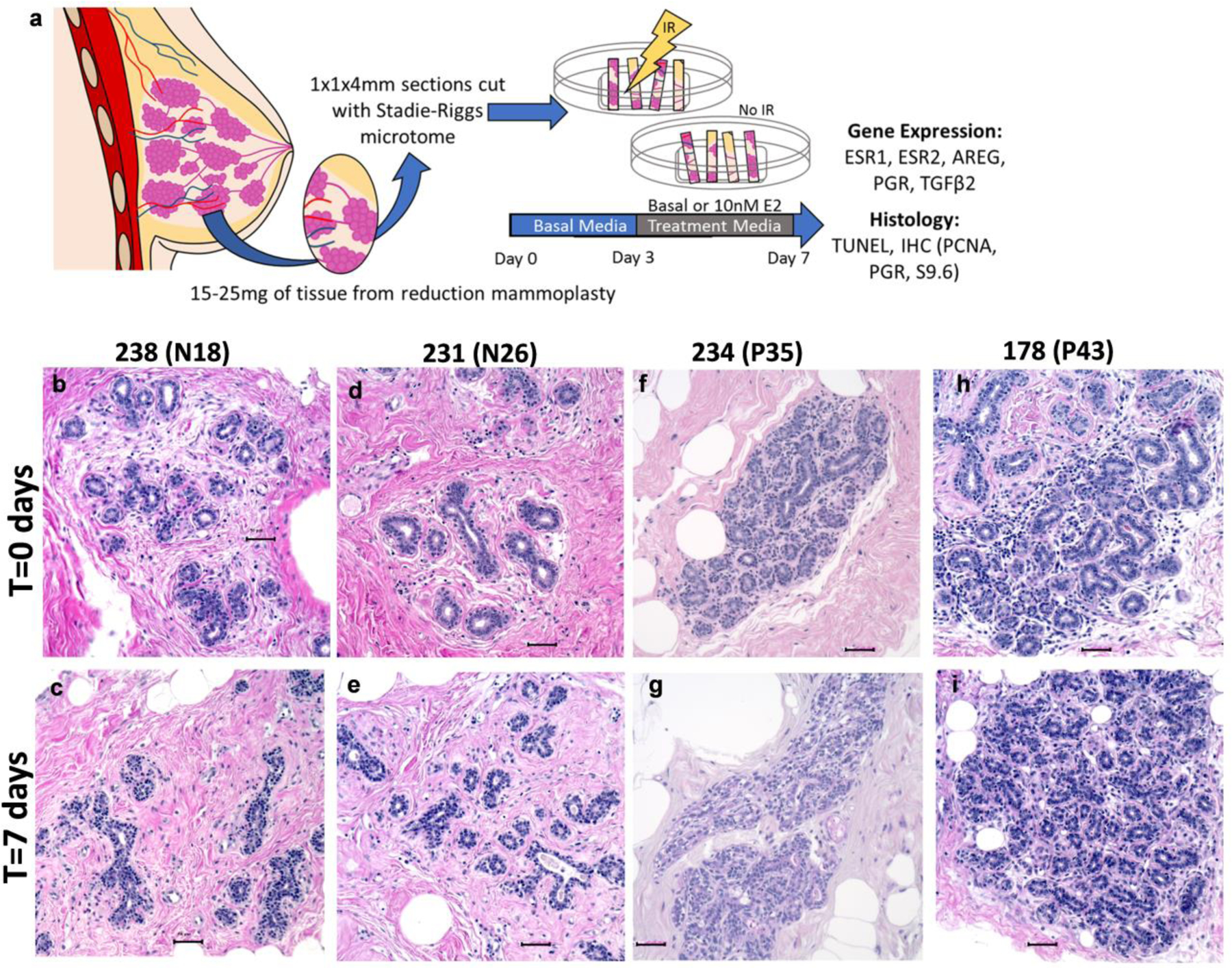
Fig. 2: Tissue architecture of human breast explants are intact after 7 days in culture.

(a) Overview of breast explant model. Tissue obtained from women undergoing reduction mammoplasties was sectioned using a Stadie-Riggs microtome. After clearing in Basal media for 3 days, explants were treated with Basal or E2 containing media for an additional 4 days. On day 7, half of the samples were subjected to 5Gy of irradiation 6 hours prior to tissue collection, and FFPE or freezing in liquid nitrogen for RNA analysis. (b-i) Representative hematoxylin and eosin staining of nulliparous (b-e) and parous (f-i) explant tissue. Comparison of fresh tissue T=0 (b,d,f,h) today 7 tissue (c,e,g,i) demonstrate that tissue architecture is maintained in culture conditions. Scale bars 50μm.
