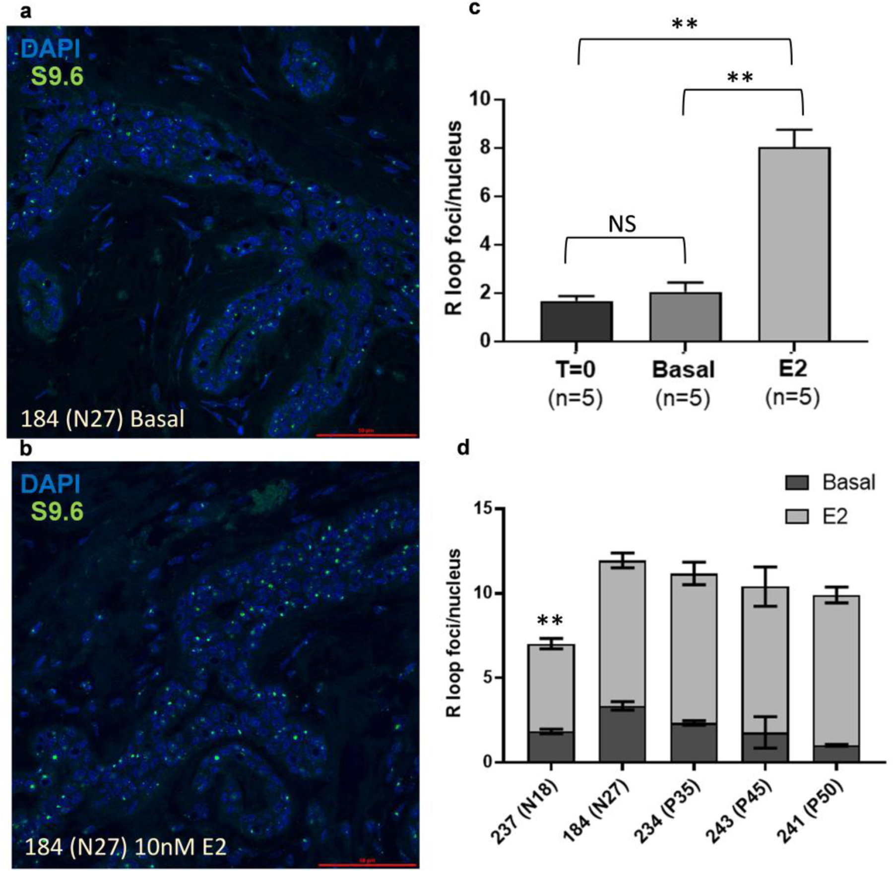
Fig. 4: E2-induces R-loop formation in human explants.

Representative images of S9.6 immunofluorescence in (a) basal and (b) E2-treated explants. (c) Quantification of R-loop foci per nucleus in basal and luminal epithelial cells demonstrates increased R-loop formation with E2-treatment relative to T=0 or basal explants (n =5 for T=0 and each treatment group). (d) R-loop foci per nucleus in Basal and 10nM E2 explants from 5 donors. Error bars indicate SEM. Significance was determined using ANOVA (** p<0.01).
