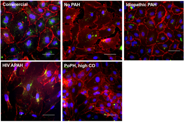
Figure 1.
Representative images confirming endothelial cell phenotype. Top left) commercial human pulmonary artery endothelial cells; passage 6. Top middle) No PAH = subject 14; passage 4. Top right) idiopathic PAH = subject 18; passage 3. Bottom left) HIV APAH = subject 13; passage 4. Bottom right) PoPH, high CO = subject 7; passage All images: VE-cadherin staining (red), acetylated low density lipoprotein uptake (green), images at 40X magnification. Scale bars = 50 μm. PAH = pulmonary arterial hypertension; HIV = human immunodeficiency virus; APAH = associated pulmonary arterial hypertension; PoPH = portopulmonary hypertension; CO = cardiac output.

