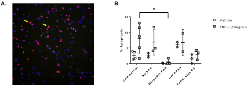
Figure 2.
Apoptosis assessed by TUNEL assay following treatment with vehicle and TNF-α. Panel A) Representative image of PAECs exposed to tert-butyl hydroperoxide for 6 hours TUNEL stained for apoptosis and counterstained with 4’,6-diamidino-2-phenylindole (DAPI). TUNEL positive cells are indicated by yellow arrow. Image at 20X magnification. Scale bar = 50 μm. Panel B) PAECs were exposed to vehicle or TNF-α for six hours. Data are presented as mean ± SD. n = 2 – 5; each n is derived from a different passage of cells from a single balloon tipped catheter from one right heart catheterization procedure for a single subject. *p < 0.05. X axis: No PAH = subject 14; idiopathic PAH = subject 18; HIV APAH = subject 13; PoPH, high CO = subject 7. PAEC = pulmonary artery endothelial cell; PAH = pulmonary arterial hypertension; PoPH = portopulmonary hypertension; CO = cardiac output.

