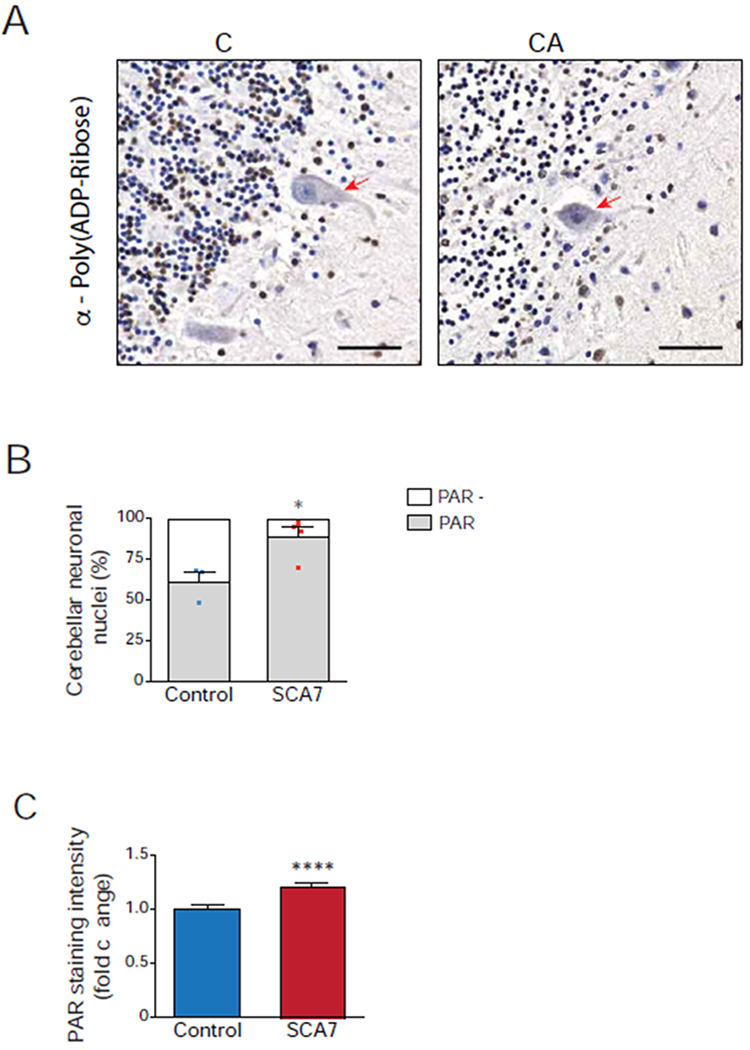
Figure 7. Human SCA7 patients display increased poly(ADP-ribose) in cerebellar neuronal nuclei.

(A) Representative images of control and SCA7 patient post-mortem cerebellar sections immunostained for poly(ADP-ribose), followed by 3,3-diaminobenzidine detection (brown) and counterstained with hematoxylin (blue). Poly(ADP-ribose)-positive nuclei stain brown, while poly(ADP-ribose)-negative nuclei stain blue, and Purkinje cells are indicated with a red arrow. Note greater fraction of cerebellar neuronal nuclei stain brown in SCA7 patient. Scale bar = 50 μM
(B) We determined the percentage of poly(ADP-ribose)-positive neuronal nuclei in cerebellar sections from four SCA7 patients and three unaffected control individuals by counting at least 300 cerebellar neuronal nuclei / individual (n ≥ 100 neuronal nuclei / section, 3 section images / individual) as either poly(ADP-ribose)-positive (brown) or poly(ADP-ribose)-negative (blue). Two-tailed t-test, *P <0.05.
(C) We quantified poly(ADP-ribose) immunoreactivity in cerebellar sections from four SCA7 patients and three unaffected control individuals by measuring 3,3-diaminobenzidine (DAB) pixel intensity in at least 300 cerebellar neuronal nuclei / individual (n ≥ 100 neuronal nuclei / section, 3 section images / individual). Two-tailed t-test, ****P <0.0001. Error bars = s.e.m.
