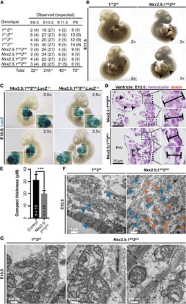
Fig. 1. Loss of Hdac1/Hdac2 within Nkx2.5IRES-Cre+ cells causes defective cardiogenesis and complete embryonic lethality.

(A) Nkx2.5IRES-Cre;1F+2F+ was crossed with 1FF2FF, and samples were recovered at various time points. aThree embryos dying. *P < 0.05; ns, not significant. (B) Nkx2.5;1KO2KO and 1FF2FF embryos at embryonic day 11.5 (E11.5) (arrows, pooled blood). (C) Nkx2.5;1Het2Het;R26R-LacZ−/+ and Nkx2.5;1KO2KO;R26R-LacZ−/+ E10.5 embryos. (D) Hematoxylin and eosin–stained Nkx2.5;1Het2Het and Nkx2.5;1KO2KO E10.5 sagittal sections at atrioventricular canal (AVC) level (arrows, eosinophilic cytoplasm; bars, compact thickness). (E) Compact myocardial thickness in control and Nkx2.5;1KO2KO E10.5 primitive ventricles (PrVs). (F) Transmission electron micrographs (TEMs) of Nkx2.5;1KO2KO and 1FF2FF E10.5 cardiomyocytes (blue, contractile fibers; orange, cytoplasmic lipid droplets). (G) TEM of Nkx2.5;1KO2KO and 1FF2FF E10.5 cardiomyocyte mitochondrial structure, density, and size.
