
Fig. 4. Hdac1 and Hdac2 cooperate to suppress cryptic transcription through deacetylation of key histone residues at alternative transcription start sites during mammalian cardiogenesis.
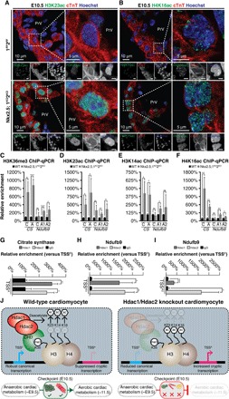
(A and B) H3K23ac (A) and H4K16 (B) staining on Nkx2.5;1KO2KO and 1FF2FF E10.5 sagittal sections at AVC level with cardiac troponin (cTnT) and Hoechst counterstains. Grayscale images are unedited. (C to E) ChIP-qPCR showing relative enrichment (TSSA versus TSSC) of Hdac1and Hdac2 at CS TSSA (C) and Ndufb9 TSSA1 (D), TSSA2 (E), normalized to immunoglobulin G (IgG). (F to I) H3K36me3 (E), H3K23ac (F), H3K14ac (G), and H4K16ac (H) ChIP-qPCR showing relative enrichment over TSSC/TSSAs for CS and Ndufb9 in Nkx2.5;1KO2KO and 1FF2FF E10.5 PHTs. (J) Hdac1/Hdac2 controls cardiomyocyte development at a critical metabolic checkpoint. CHD, chromodomain containing subunit. WT, wild type.