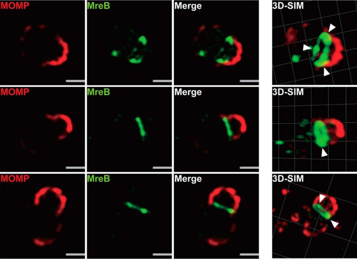
FIG 1.
Localization of chlamydial MreB_6×H in C. trachomatis using structured illumination microscopy (SIM). C. trachomatis without plasmid (−pL2) was transformed with an anhydrotetracycline (aTc)-inducible vector encoding chlamydial MreB with a six-histidine (6×H) tag at the C terminus. HeLa cells were infected with this strain and chlamydial MreB_6×H expression was induced with 10 nM aTc at 6 hpi. At 10.5 hpi, the infected cells were fixed (3.2% formaldehyde, 0.022% glutaraldehyde in PBS) for 2 min and permeabilized with 90% methanol (MeOH) for 1 min. The sample was stained for major outer membrane protein (MOMP; red) and chlamydial MreB (green). Three representative images are displayed. The arrowheads indicate regions of more intense fluorescence. SIM images were acquired on a Zeiss ELYRA PS.1 superresolution microscope. Bars, 0.5 μm.

