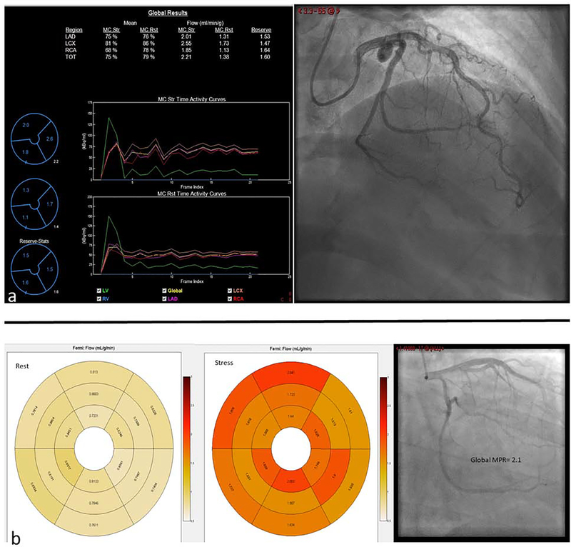
Central Illustration: Abnormal MPR in non-obstructive coronary artery disease.
PET quantification of MBF shows reduced MPR of 1.6 in this 48 year old female with non-obstructive CAD (a). Rest and stress CMR derived segmental quantification of myocardial blood flow with a global MPR of 2.1 in a 59 year old male with non-obstructive CAD. LAD-left anterior descending artery, LCX-left circumflex artery, RCA-right coronary artery, TOT-total, MC-motion corrected, Str-Stress, Rst-Rest (b).

