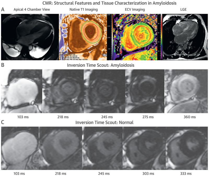
FIGURE 4. CMR in CA.
Cardiovascular magnetic resonance (CMR) provides characteristic imaging of the (A) structural changes and (B) powerful tissue characterization with features of high native T1, expanded extracellular volume (ECV), and late gadolinium enhancement (LGE) (diffuse, subendocardial, or transmural). (C) Post-gadolinium myocardial signal intensity changes characteristically with myocardial signal nulling before the blood pool signal in amyloidosis and vice versa in non-amyloid hearts. Abbreviation as in Figure 1.

