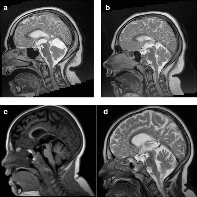
Fig. 1.
a MRI image of patient 1 (family 1), sagittal section. In T2-weigted image, thinning of the posterior part of corpus callosum is visible as well as an infratentorial cyst modeling the cerebellum. See also brachycephaly due to small brain. b MRI image of patient 2 (family 1), sagittal section. In T2-weighted image, thinning of a posterior part of corpus callosum is visible. See also brachycephaly due to small brain. c, d MRI of patient 3 (family 2), sagittal section. In T1-weigted image, the similar thinning of the posterior part of corpus callosum is visible. In this patient, a relative white matter loss can also be recognized in a T2 sagittal section

