Figure 2.
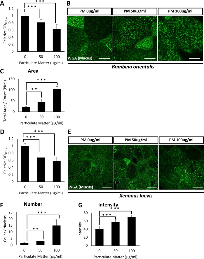
Particulate matter from heavy diesel engine disturbs mucus secretion from the mucociliary epithelium in B. orientalis and X. laevis embryos. (A) Increasing doses of PM were administered to B. orientalis embryos and secreted mucus levels were analyzed by ELLA using WGA-HRP. 50 µg/mL or 100 µg/mL of PM significantly disturbed mucus secretion from the embryonic epidermis. Statistical analysis was performed using one-way ANOVA. From left to right, n = 21, 16, 20. (B) The WGA positive mucus vesicles were visualized by fluorescent microscopy. PM exposure significantly accumulated mucus vesicles in the cytoplasm. Scale bar = 10 μm. (C) Quantification of WGA positive mucus vesicles in (B). Statistical analysis was performed using one-way ANOVA. From left to right, n = 4, 4, 4. (D) Particulate matter-induced hyposecretion of mucus was recovered by α-tocopherol in Xenopus embryos. Statistical analysis was performed using one-way ANOVA. From left to right, n = 24, 22, 22. (E) The WGA positive mucus vesicles were visualized by fluorescent microscopy. The exposure to the particulate matter significantly accumulated the mucus vesicles in the cytosol. Scale bar = 20 μm. (F,G) Quantification of WGA positive mucus vesicles in (E). For (F), statistical analysis was performed using one-way ANOVA. From left to right, n = 4, 4, 4. For (G), statistical analysis was performed using one-way ANOVA. From left to right, n = 30, 30, 30. Asterisks represent ***(p < 0.001), **(0.001 < p < 0.01), *(0.01 < p < 0.05), ns (0.05 < p). PM, particulate matter; ELLA, enzyme-linked lectin assay.

