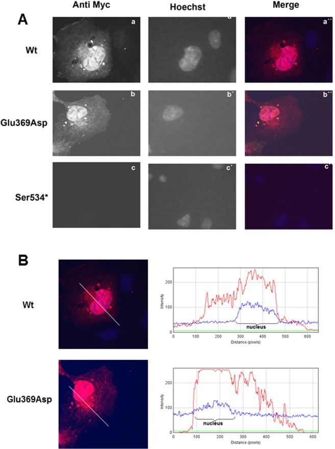
Figure 5.
(A) Expression pattern of the EYA4-HR Wt (a), Glu369Asp (b) and Ser534* (c) in COS7 cells. EYA4-HR protein shows a cytoplasmic and nuclear distribution for the Wt and Glu369Asp mutant but no signal was observed for the mutant Ser534*. Hoechst staining was used to identify the nucleus. (B) IMAGE J graphical section showing the fluorescence intensities plotted against the distances (pixels). EYA4-HR staining (in red) for the Wt and Glu369Asp mutant is detected in both the nucleus and the cytoplasm.

