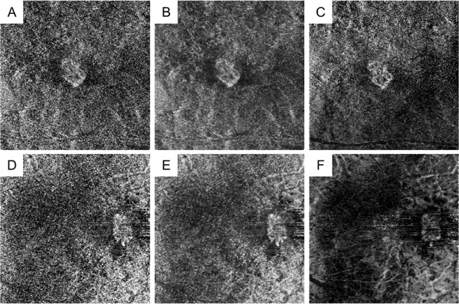
Figure 1.
Representative optical coherence tomography angiography (OCTA) images of myopic choroidal neovascularisation (mCNV) after denoising and averaging processes. Images of a 76-year-old female patient (A–C) and a 60-year-old female patient (D–F) with mCNV in the right and left eyes, respectively. (A,D) Single OCTA images before the denoising process are shown. (B,E) Single OCTA images after the denoising process are shown. (C,F) OCTA images after the averaging process with a maximum of 10 single images are shown. The denoising process allowed for mCNV visualisation with a high contrast.

