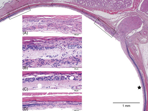
Figure 16.24.

Light-induced retinal degeneration in an albino rat from a 2-year carcinogenicity study. There is a distinct difference in thickness of the retina on either side of the optic disk. The ★ marks the relatively normal inferior retina. Approximately 50% of the superior retina is markedly reduced in thickness and shows different degrees of retinal atrophy. These begin adjacent to the disk (boxed area-.A, Figure 16.24A) and extend to the midperiphery (boxed area-D, Figure 16.24D); intervening regions are illustrated in 16.24B and C. (A) The retina nearest the optic disk shows full-thickness loss of retinal layer organization and fibrous connective tissue in the vitreal aspect of degenerating retina. (B) The same tissue overlays the ganglion cell layer; the inner nuclear layer is moderately reduced and abuts the choroid and sclera. The outer retinal layers and RPE are absent. (C) Microcystoid vacuoles are present within remnant inner retinal layers. (D) There is an abrupt transition into less affected retina with preservation of RPE, outer nuclear, and photoreceptor layers.
de Vera Mudry et al. (2013); reprinted by permission of Sage Publications.
