
Fig. 10.
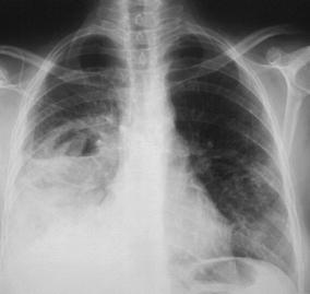
Typical radiographic characteristics of acute lobar pneumonia in a 53-year-old inpatient. The chest radiogram displays an extensive, dense alveolar infiltrate in the right mid- and lower lung fields with loss of the silhouettes of the lower right heart border and right hemidiaphragm. These features suggest involvement of the right lower lobe and middle lobe by the pneumonic infiltrate. In addition, an air–fluid level indicates cavitation. Note that the infection has spread transbronchially to the left lung. The patient was diagnosed to have E. coli pneumonia