
Fig. 12a,b.
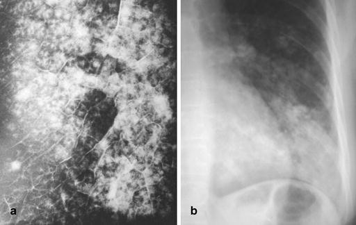
Radiographic appearance of lobular (broncho) pneumonia. a Specimen radiogram of lobular (broncho) pneumonia. This radiogram, obtained from a post-mortem examination of a patient with Staphylococcus aureus pneumonia, demonstrates the centrilobular origin of this particular form of pneumonia. As disease spreads from the walls of terminal and respiratory bronchioles, large portions of secondary lobules are involved, and confluent disease may cause frank lobar consolidation. [By courtesy of Saunders, Philadelphia. Potchen J, Grainger R, Greene R (eds) Pulmonary radiology.] b Radiographic appearance of lobular (broncho) pneumonia caused by Staphylococcus aureus and shows patchy and partly confluent densities in the left lower lobe. This pattern is distinctly different from the one seen in acute lobar (air-space) pneumonia