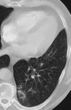
Fig. 13.

Computed tomography findings in early bronchopneumonia. The CT section through the left lower lobe in a patient with Haemophilus influenzae pneumonia demonstrates small nodular and patchy acinar lesions with a tendency towards confluence in the posterior basal segment of the left lower lobe
