
Fig. 15.
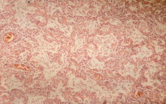
Histopathologic correlate of interstitial pneumonia. This histopathologic specimen (hematoxylin–eosin stain) demonstrates moderate lymphocytic infiltrates surrounding a blood vessel and extending into the neighboring alveolar walls. The presence of macrophages and pneumocytes suggest the beginning of acute alveolar damage