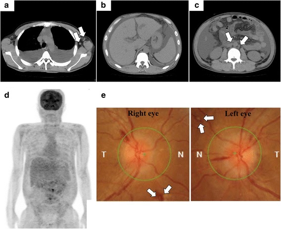
Fig. 1.

Imaging findings. a–c Computed tomography images on the day of admission. a Massive pleural effusion and slightly enlarged axillary lymph nodes are observed (arrows). b Hepatosplenomegaly is seen. c Massive ascites and slightly enlarged para-aortic lymph nodes are observed (arrows). d 18F–fluorodeoxy-glucose positron emission tomography (FDG-PET) images on day 8. Although the findings are poor (FDG uptake is generally weak), FDG uptake is observed in the para-aortic lymph nodes. e Funduscopic evaluation performed on day 10. Bilateral optic disk edema is remarkable. Roth’s spots are observed (arrows). Hemorrhage in the fundus of right eye is also observed
