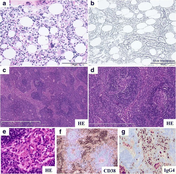
Fig. 2.

Pathological findings. a, b Pathological evaluation involving bone marrow biopsy in the present case performed on day 9. a Mild increased number of megakaryocytes is observed (hematoxylin and eosin [H&E] staining). b Mild reticulin myelofibrosis in bone marrow is observed (silver impregnation staining). c–h Pathological evaluation of the right neck lymph node in the present case. c, d Atrophic germinal center, vascular invasion with a glomerular-like pattern of vascular endothelial cell proliferation and hyalinization are observed in the follicles (H&E staining). e Dendric proliferation of arterioles and swelling of vascular endothelial cells are observed (H&E staining). f, g Invasion of plasma cells (CD38+) is observed in the intrafollicular space (immunohistochemical staining). h Immunoglobulin G (IgG) 4-positive plasma cells are observed (> 10 IgG4-positive plasma cells/high power field on immunohistochemical staining); however, the IgG4/IgG ratio is 24.2%, and it does not fulfill the criteria for IgG4-related disease (> 40%, data not shown). Human herpesvirus 8 is negative on immunohistochemical staining, and the Epstein-Barr virus-encoded small RNA in situ hybridization is negative in the lymph node (data not shown). These findings are compatible with mixed-type multicentric Castleman disease-like histology
