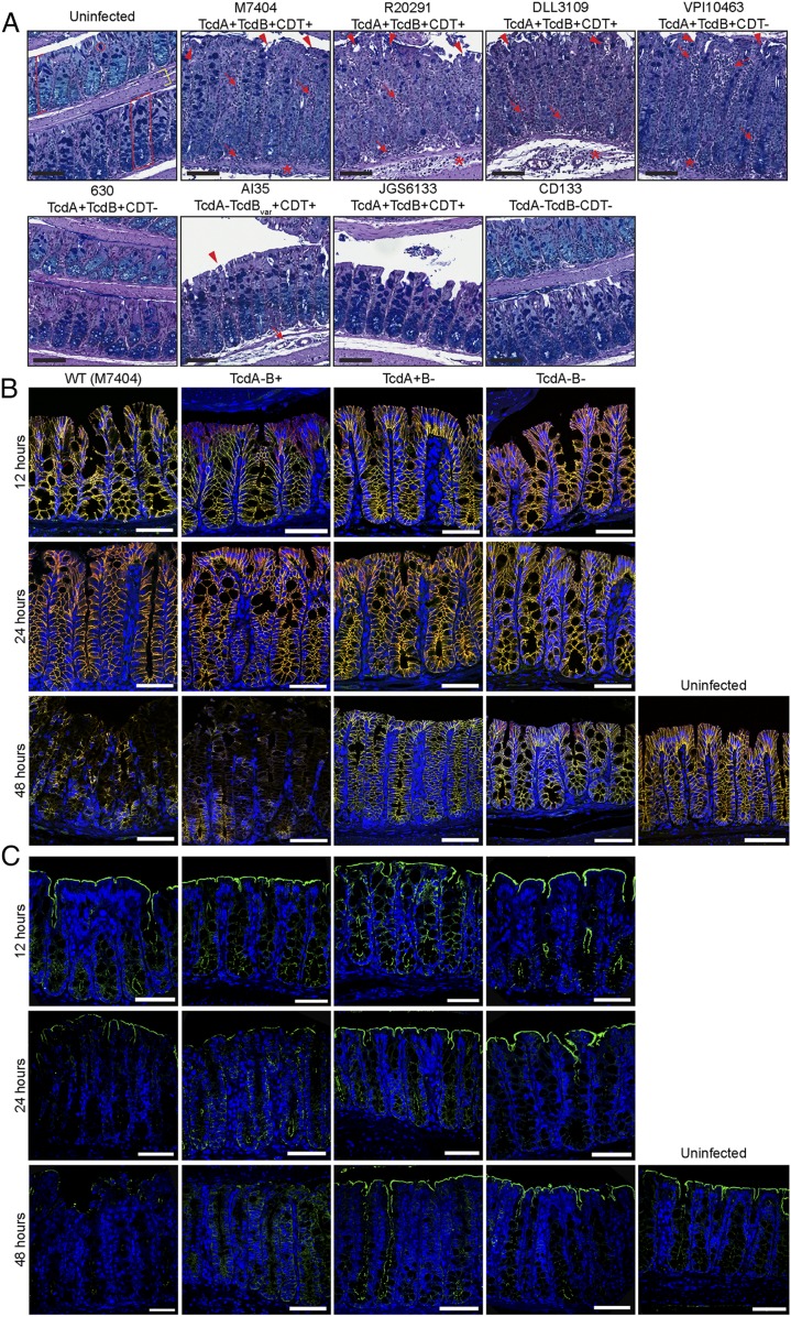
Fig. 1.
C. difficile induces severe and deep epithelial damage through TcdB alterations in adherens-junction formation and cellular polarity. (A) Mice were infected with a panel of genetically distinct C. difficile isolates and monitored for disease severity and colonic damage through periodic acid–Schiff (PAS)/Alcian blue staining. Representative images of swiss-rolled colonic tissue are shown. The colonic mucosa (red bracket) and, more specifically, the crypts of Lieberkühn (red box), comprised of colonic epithelial cells and goblet cells (red circle), sit above the submucosa and muscle layers (yellow bracket) of the colon. Arrow, inflammation; arrowhead, crypt damage/goblet cell loss; asterisk, edema. n ≥ 5. (Black scale bars: 100 µm.) (B and C) Colonic tissues were collected at 12, 24, and 48 h postinfection with M7404 (WT), and the isogenic toxin mutants of this strain (TcdA−B+, TcdA+B−, and TcdA−B− C. difficile), or from uninfected mice (48 h). A representative image of tissues stained for (B) β-catenin (green) and E-cadherin (red) (merge, yellow) or (C) ezrin (green) is shown, with nuclei stained with DAPI (blue). n ≥ 5. (White scale bars: 50 µm.) See also SI Appendix, Fig. S1.

