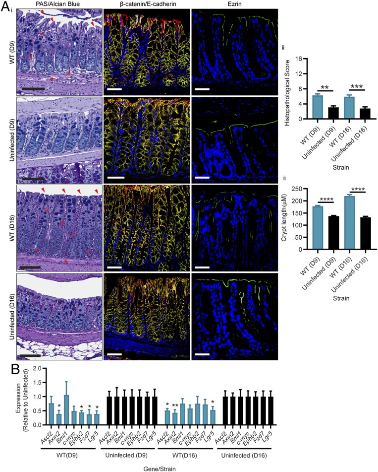
Fig. 4.
C. difficile TcdB-mediated stem cell death and dysfunction hinders host repair for up to 2 wk. Mice were infected with M7404 (WT) C. difficile or left uninfected and allowed to recover after reaching a peak of infection (between 10% and 15% weight loss). Two weeks post the peak of infection, (A) colonic tissues were collected and assessed with (i) PAS/Alcian blue for overall pathology, β-catenin (green), and E-cadherin (red) (merge, yellow) for adherens junctions, or ezrin (green) for cellular polarity. (ii) Overall histopathology was scored and plotted, and (iii) crypt length was measured for 30 crypts per mouse across the entire length of the colon. Arrows, inflammation; arrowheads, crypt damage/goblet cell loss; asterisk, edema. (Scale bars: black, 100 µm; white, 50 µm.) (B) Quantitative ddPCR analysis of colonic tissue 9 d (D9) and 16 d (D16) postinfection (7 and 14 d post peak of infection, respectively). Gene expression, as fold change relative to uninfected mice, was plotted. n ≥ 5. Data are represented as mean + SEM. *P ≤ 0.05, **P ≤ 0.01, ***P ≤ 0.001, ****P ≤ 0.0001.

