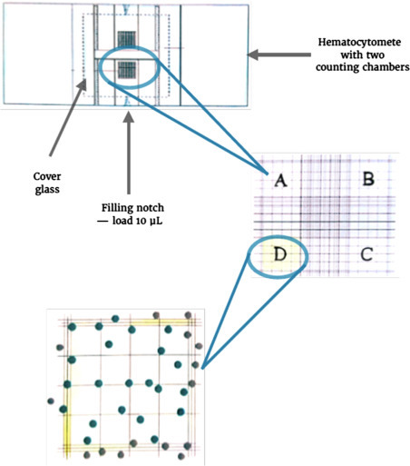
Figure 9.4.

Cell quantification using Trypan Blue.
The hemacytometer is prepared by covering both counting chambers with a cover glass. Subsequently, 10 μL of a 1:1 cell suspension with 0.4 % Trypan Blue is loaded onto the filling notch of one of the counting chambers. Through capillary action, this volume will cover the grid that can be observed in an inverted microscope at magnifications of at least 10X. The average of cells covering squares A–D determines the number of cells per mm2. Viable cells in these squares are counted by excluding nonviable cells that appear black due to their absorption of Trypan Blue through their permeable cell membranes. Only cells overlapping with one of the outer horizontal and vertical borders should be included.
