Fig. 1.
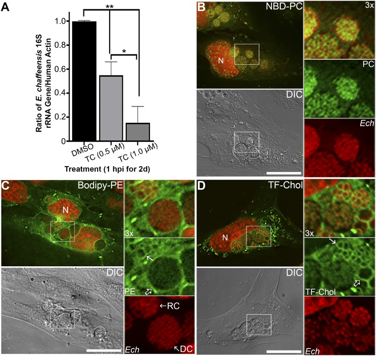
E. chaffeensis is dependent on host-derived lipids and incorporates exogenous phospholipids and cholesterol. (A) E. chaffeensis-infected THP-1 cells were seeded in a six-well plate. At 1 hpi, cells were incubated with 0, 0.5, or 1 μM triacsin C (TC) for 2 d at 37 °C. DNA was extracted from treated samples, and quantitative PCR was performed for the E. chaffeensis 16S rRNA gene and normalized against human ACTIN. Results are shown as the mean ± SD from three independent experiments. **P < 0.001; *P < 0.01 (ANOVA). (B–D) RF/6A cells were seeded onto cover glasses in a six-well plate for 3 h and then infected with E. chaffeensis (Ech). Cells were incubated with 25 μM NBD-PC for 1 d at 1 dpi (B), or with 5 μM Bodipy-PE for 4 h at 2 dpi (C); Alternatively, infected cells at 1 dpi were washed and replaced with AMEM containing lipoprotein-depleted serum for 8 h, then incubated with 1 μM TF-Chol for 1 d (D). Cells were fixed and DNA was stained with Hoechst 33342 to label host and bacterial DNA (pseudocolored in red). Samples were observed under a DeltaVision microscope. The boxed area in the merged image is enlarged 3× on the Right. DC, dense-core forms with diameter <1 μm and tightly packed chromosomes; DIC, differential interference contrast; RC, reticulate cell forms of E. chaffeensis with larger diameter (≥1 μm) and more loosely-packed chromosome DNAs. Open arrow, inclusion membrane; solid arrow, E. chaffeensis membrane; N, nucleus. Images are representative of at least three independent experiments. (Scale bars, 10 μm.)

