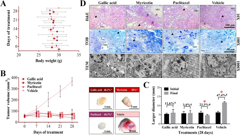
Fig. 4.
Antineoplastic activity of GA and Myr in mice xenotransplanted with ovarian cancer. The body weight of rodents was monitored with an electronic bascule for 28 days (a). The tumor volume was determined with a Vernier caliper in mice treated with GA and Myr for 4 weeks, with doses of 50 mg/kg in 2 alternate days per week by peritumoral route (Tumoral volume = [Larger diameter * (Shorter diameter)2] / 2). Additionally, morphological changes in tumor lesions were analyzed at the end of the treatments, and the % inhibition was calculated (b). Subsequently, the disease evolution was evaluated in each treatment with the larger diameter obtained in tumoral lesions at the beginning and final of the assay. Also, the % inhibition of tumoral volume was determined based on the final volume of each tumor after treatment concerning the final volume obtained by the control group (c). Differential histological patterns were observed in the tumor lesions by H&E / TOB stain and by TEM (d). Results show the mean ± S.D. of two biological replicates (n = 5); *, p ≤ 0.05 vs. the control group without treatment (20 μL of 0.5% DMSO in 1X PBS, ANOVA). Paclitaxel was used as a positive control at 5 mg/kg body weight, administered under the same conditions that experimental samples. The arrow’s direction indicates gain (↑) or loss (↓) of tumoral volume (c). The arrows and symbols indicate: fibrosis (black arrowhead), necrotic area (NEC), vascularization (*), leukocytic infiltrates (grey arrowhead), and apoptotic cells (white arrowhead) (d). H&E, hematoxylin and eosin; TOB, toluidine blue; TEM, transmission electron microscopy

