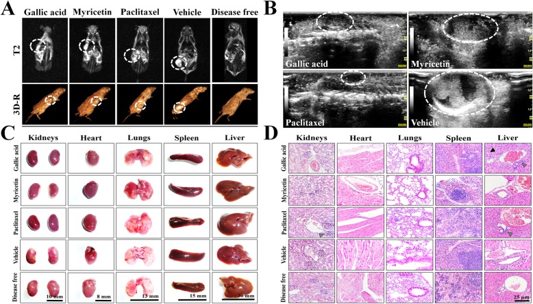
Fig. 5.
Imaging and histopathologic studies in mice treated with GA and Myr.NMR (a) and USG (b) were performed to observe densitometric and morphological changes in the tumoral lesions, as well to discard metastatic processes during treatments. The tumor lesions were delimited with a white circle in the corresponding images of both studies. Observations of the anatomical morphology (c) and the histological patterns (d) of organs extracted after laparotomy were made, to discard tissue lesions caused by treatments (50 mg/kg in 2 alternate days per week, 4 weeks, peritumoral route). Histopathology images were taken at 40X magnification and color arrows indicate loss of hepatic parenchyma (black arrowhead) or leukocyte infiltrates (grey arrowhead). Results are representative of two biological replicates (n = 5). NMR, nuclear magnetic resonance; T2, transverse relaxation times; 3D-R, 3D-reconstruction; USG, ultrasonography

