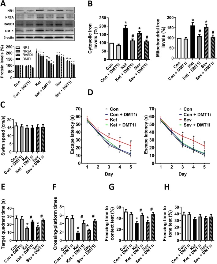
Fig. 6.
GA-induced activation of NMDAR-RASD1-mediated DMT1 iron uptake signalling pathway. a To inhibit NMDAR-RASD1-mediated DMT1 iron uptake signalling pathway, DMT1i was added to the culture medium 30 min before GA exposure. Protein levels of NMDAR, RASD1, and DMT1 were determined by Western blot. A representative image set is presented. b The cytosolic and mitochondrial iron levels of hippocampal neurons. c Swim speed, d escape latency, e time spent in the target quadrant, and f crossing platform times in MWM tests. g Freezing time to context and h freezing time to tone in the fear conditioning tests. a and b for hippocampal neurons; c–f for adolescent rats; g and h for aged mice. Data are presented as mean ± SEM (n = 12 animals/group); *p < 0.05 compared with Con group; #p < 0.05 compared with the GA (Ket or Sev) group

