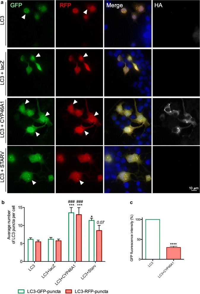
Fig. 2.
CYP46A1 overexpression significantly increases the number of LC3 puncta upon autophagy inhibition. a N2a cells were transfected with ptfLC3-RFP-GFP, co-transfected with ptfLC3-RFP-GFP and CYP46A1, and co-transfected with ptfLC3-RFP-GFP and lacZ. An additional condition was used, promoting starvation, as a positive control for autophagy activation. The visualization of LC3-puncta is possible upon autophagy inhibition with chloroquine in all the experimental conditions. The presence of LC3-RFP-GFP puncta (co-localized) refers to the presence of mature autophagosomes, whereas LC3-RFP puncta (without GFP) refers to autolysosomes. Representative microscopy images. b For each condition 100 cells were randomly counted in different microscopy fields. The number LC3-puncta per cell upon CYP46A1 overexpression was significantly increased compared to both control conditions. c The total fluorescence intensity (GFP) was significantly reduced upon CYP46A1 overexpression, compared to control conditions, thus suggesting an increase in the autophagic clearance. Values expressed as mean ± SEM. (a, b, n = 5 independent experiments, two-way ANOVA with Bonferroni’s multiple comparisons test, *P < 0.05; ***P < 0.0001;###P < 0.0001 comparing to LC3 + lacZ; c, n = 3 independent experiments Unpaired Student’s t test, ****P < 0.00001)

