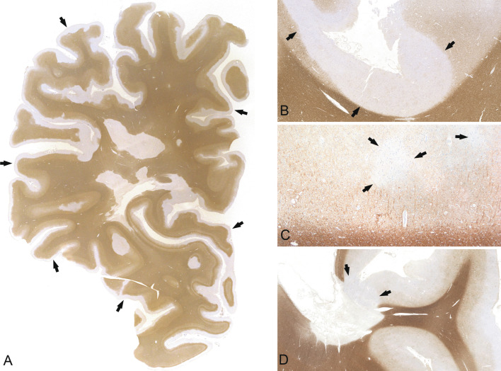
Fig. 19.6.
Cortical demyelination. Cortical demyelination located subpially (A, B), intracortically (C), and overlapping with the white matter (so-called compound plaques) (D). A subpial lesion extends over several gyri and sulci and covers almost the entire cortex (A, proteolipid protein, arrows; B, myelin basic protein (MBP), arrows). A small, demyelinated lesion is located intracortically (C, MBP, arrows). A compound plaque is overlapping the cortex and the subcortical white matter (D, MBP, arrows). B, × 1.56; C, × 40; D, × 0.59.

