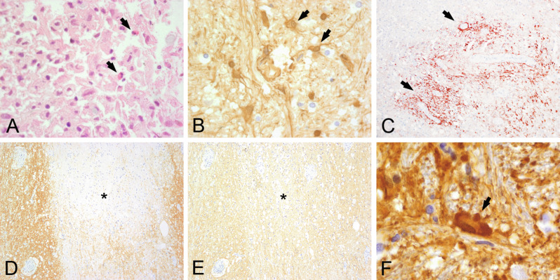
Fig. 19.9.
Active stage of neuromyelitis optica (NMO) lesion. An active NMO lesion shows inflammatory infiltrates with eosinophils (A, hematoxylin and eosin) next to macrophages and lymphocytes, deposition of immunoglobulin G (B, IgG), and C9 neoantigen (C, arrows). Some lesions show selective loss of aquaporin-4 (D, asterisk marks lesion) while aquaporin-1 is well preserved (E) and there is accumulation of bizarre astrocytes with beading and clumping of cell processes (clasmatodendrosis) (F, glial fibrillary acidic protein). A, B, F, × 600; C, × 200; D, E, × 100.

