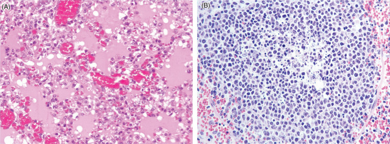
Figure 8.7.

African swine fever virus infection in a domestic pig.
(A) Diffuse pulmonary edema with flooding of alveolar air spaces by edema and foamy macrophages. There is hemorrhage within alveolar septae. (B) Necrosis of lymphocytes within a lymph node with central cellular debris and apoptotic bodies. Surrounding the follicle are extravasated erythrocytes.
(Photos Courtesy of the Department of Compared Anatomy and Anatomic Pathology. Veterinary Faculty of the University of Cordoba, Spain [Archive].)
