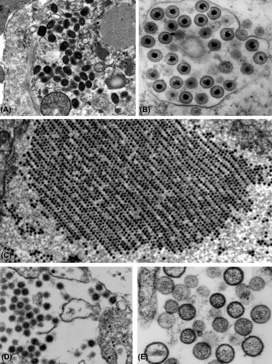
Figure 1.3.

Thin-section electron microscopy of selected viruses. The remarkable diversity of the viruses is clearly revealed by thin-section electron microscopy of infected cells—and this technique provides important information about morphogenesis and cytopathology. (A) Family Poxviridae, genus Orthopoxvirus, variola virus. (B) Family Herpesviridae, genus Simplexvirus, human herpesvirus 1. (C) Family Adenoviridae, genus Mastadenovirus, human adenovirus 5. (D) Family Togaviridae, genus Alphavirus, Eastern equine encephalitis virus. (E) Family Bunyaviridae, genus Hantavirus, Sin Nombre virus. These images represent various magnifications; the details of the morphogenesis of the various viruses are given in the chapters of Part II of this book.
