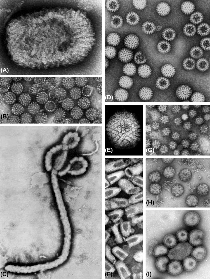
Figure 1.4.
Negative contrast electron microscopy of selected viruses. The remarkable diversity of the viruses is revealed by all kinds of electron microscopy methods, but none better than by negative staining. (A) Family Poxviridae, genus Orthopoxvirus, vaccinia virus. (B) Family Papovaviridae, genus Papillomavirus, human papillomavirus. (C) Family Filoviridae, Ebola virus. (D) Family Reoviridae, genus Rotavirus, human rotavirus. (E) Family Herpesviridae, genus Simplexvirus, human herpesvirus 1 (capsid only, envelope not shown). (F) Family Rhabdoviridae, genus Lyssavirus, rabies virus. (G) Family Caliciviridae, genus Norovirus, human norovirus. (H) Family Bunyaviridae, genus Phlebovirus, Rift Valley fever virus. (I) Family Orthomyxoviridae, genus Influenzavirus A, influenza virus A/Hong Kong/1/68 (H3N2). These images represent various magnifications; the size of the various viruses is given in Chapter 2: Classification of Viruses and Phylogenetic Relationships and in the chapters of Part II of this book.

