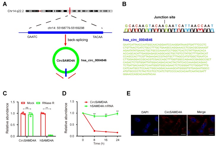
Figure 2.
Characterization of circSAMD4A in preadipocytes. (A) Genomic location of the hSAMD4A gene and of circSAMD4A. (B) Sanger sequencing showing the “head-to-tail” splicing of circSAMD4A in preadipocytes. (C) qRT-PCR of quantification of circSAMD4A and hSAMD4A mRNA expression in preadipocytes after treatment with RNase R. (D) qRT-PCR quantification of circSAMD4A and hSAMD4A mRNA expression in preadipocytes after treatment with Actinomycin D. (E) RNA FISH for circSAMD4A. Nuclei were stained with DAPI. Scale bar = 20µm. Data are presented as means ± SD; significant difference was identified with Student's t test. *P < 0.05; **P < 0.01; ns (not significant).

