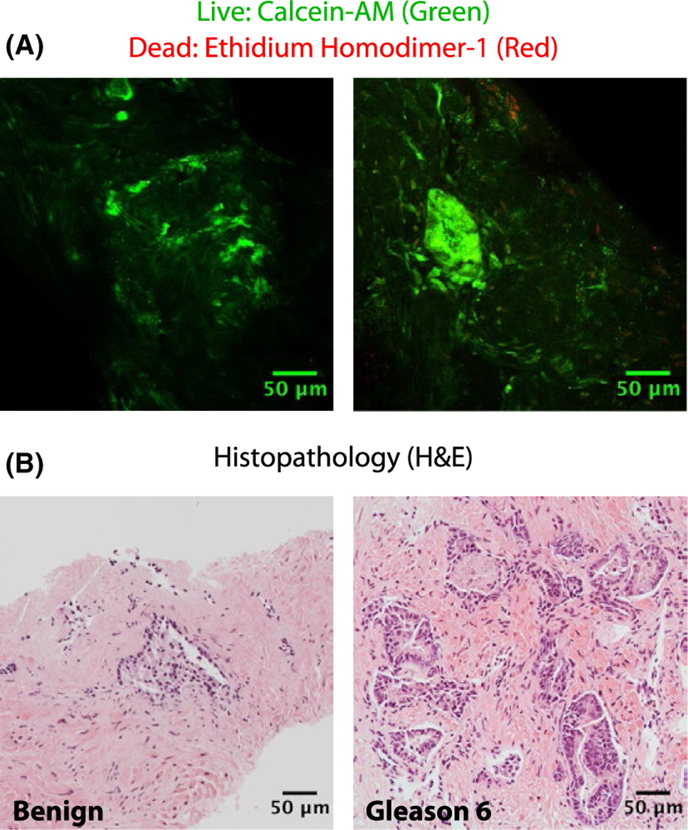
FIGURE 6.

Biopsy LIVE/DEAD staining. (A) Confocal 2-photon microscopy from biopsy samples stained with calcein-AM and ethidium homodimer-1 following 26 h in culture. (B) Biopsy histopathology obtained after confocal microscopy. Each hematoxylin and eosin (H&E) stained image originates from the same sample as the confocal image directly above but not from the exact same location in the tissue
