Figure 1.

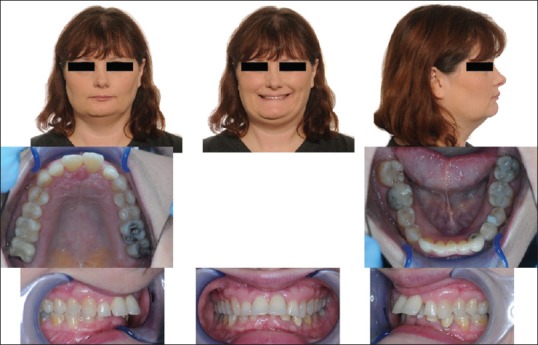
Pre-treatment clinical images. Facial photographs show repose and smile views. Clinical intra-oral images illustrate excessive horizontal and vertical overlaps of the dentition, consistent with Class II Division 1 malocclusion

Pre-treatment clinical images. Facial photographs show repose and smile views. Clinical intra-oral images illustrate excessive horizontal and vertical overlaps of the dentition, consistent with Class II Division 1 malocclusion