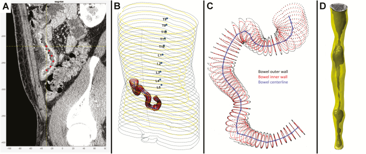
Figure 1.
Illustration of bowel segmentation and reconstruction process. A, Radiologist places reference center points to identify distal small bowel. B, Centerline within small bowel is shown in 3D space in the context of patient abdominal silhouette. C, Centerline shown with automatically identified inner and outer bowel wall perimeters using superpixel voxel segmentation followed by k-means classification, allowing segmentation of lumen, bowel wall, and extra-intestinal space. D, Example of straightened tube-like reconstruction of segmented intestine with bowel wall and shaded lumen identified.

