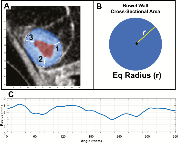
Figure 3.
Comparing linear on-screen measurements and automated equivalent linear measures derived from cross-sectional areas. A, An example axial section of diseased small bowel with bowel wall masked in blue and lumen masked in red based on automated segmentation. B, The process of equivalent radius generation, where the total bowel wall area is reformed into a perfect circle and the (equivalent) radius is measured as the square root of Area/ π. In this case, the equivalent BWT radius is 7.3 mm compared with radiologist A measuring 8.1 mm and radiologist B measuring 7.1 mm. C, The variability of bowel wall thickness over 360 degrees of rotation about the estimated lumen center. Measures vary between a bowel wall thickness of 8.3 mm and 4.1 mm based on the location of measurement in the same cross-sectional segment.

