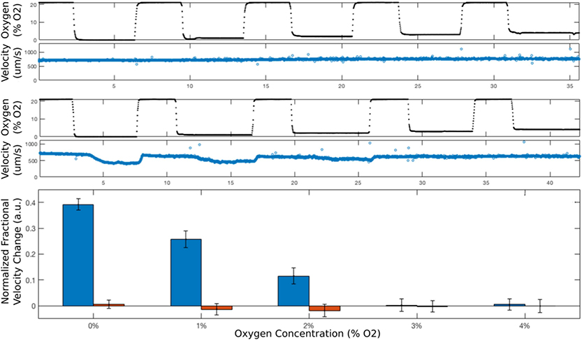
Figure 2. Flow velocity of SCT blood is oxygen-dependent at low oxygen.
(Top) For a normal blood sample (AA) containing only HbA, when the oxygen concentration (black dots in top panel) is cycled between 21% and low concentrations, first 0%, then 1%, 2%, 3%, 4%, the flow velocity (blue circles in the second panel from the top) does not change. In contrast, when the same oxygen concentration cycle (black dots in the third panel from the top) is applied to a flowing SCT blood sample (36% HbS), the flow velocity (blue circles in fourth panel from the top) drops at the lower oxygen concentrations because of sufficient hemoglobin polymerization and RBC sickling. The bottom panel compares the relative velocity changes of SCT and AA blood and shows that hemoglobin polymerization and RBC sickling are sufficient to reduce SCT flow velocity at oxygen concentrations at least as high as 2%. Error bars indicate one standard deviation.

