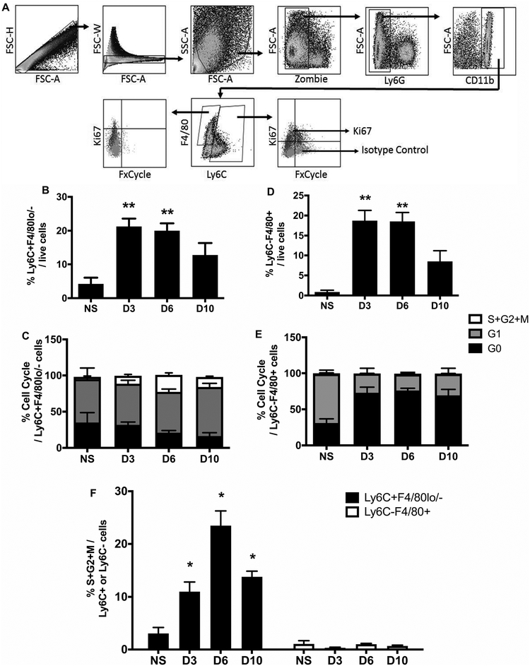
Figure 2. Skin wounding increases proliferation of Ly6C+ Mo/MΦ but not mature Ly6C- MΦ.
(A) Representative flow cytometric images showing proliferative profiles of Ly6C+F4/80lo/- Mo/MΦ and Ly6C-F4/80+ MΦ in skin wounds on day 6 post-injury. (B) and (D) Percentages of Ly6C+F4/80lo/- Mo/MΦ and Ly6C-F4/80+ MΦ of total live cells in normal skin (n=4) and wounds on days 3 (n=6), 6 (n=9), and 10 (n=7) post-injury. (C) and (E) Percentages of Ki67-FxCycle- cells (G0 phase, black), Ki67+FxCycle- cells (G1 phase, grey), and Ki67+FxCycle+ cells (S/G2/M phases, empty) in Ly6C+F4/80lo/- Mo/MΦ and Ly6C-F4/80+ MΦ. (F) Comparison of Ki67+FxCycle+ cells (S/G2/M phases) in Ly6C+F4/80lo/- Mo/MΦ (black bars) and Ly6C-F4/80+ MΦ (open bars) during wound healing. For each graph, data were pooled over three separate time course experiments; sample sizes given are pooled over experiments. Data expressed as mean ± SEM; *P < 0.05 or **P < 0.01 vs non-injured skin group by ANOVA.

