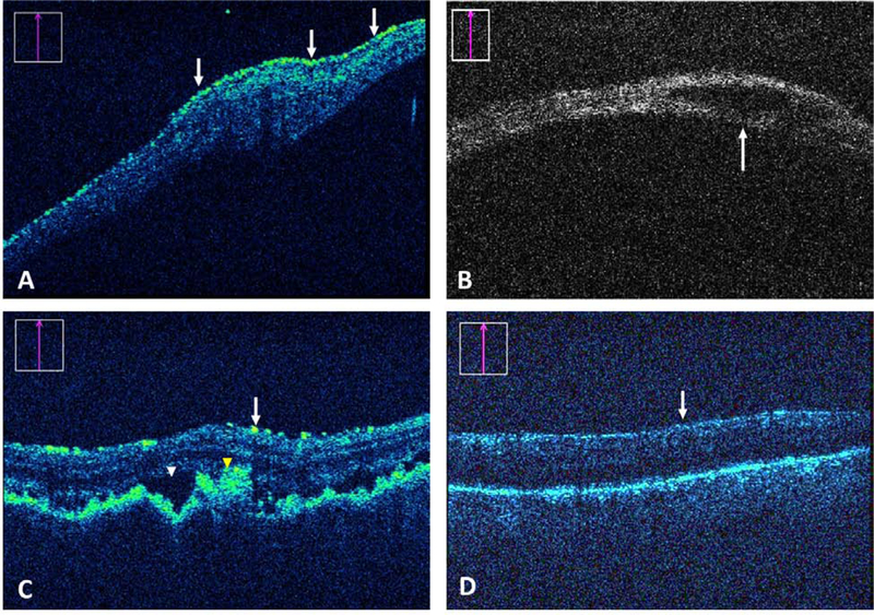
Figure 2.

Impact of iOCT on surgical decision-making. (A) Preretinal membrane (arrows) identification by iOCT that prompted further peeling. (B) iOCT-based identification of a retinal cyst (arrow) confirming no need for additional treatment. (C) A complex combined exudative/rhegmatogenous detachment repair with suspected uveitis and choroidal folds in which SRF (white arrowhead) and abnormal pigment (yellow arrowhead) were identified by iOCT. The feedback resulted in more drainage. (D) In a possible proliferative vitreoretinopathy case, iOCT confirmed lack of preretinal membranes (arrow) that prevented unnecessary staining or membrane peel attempts.
