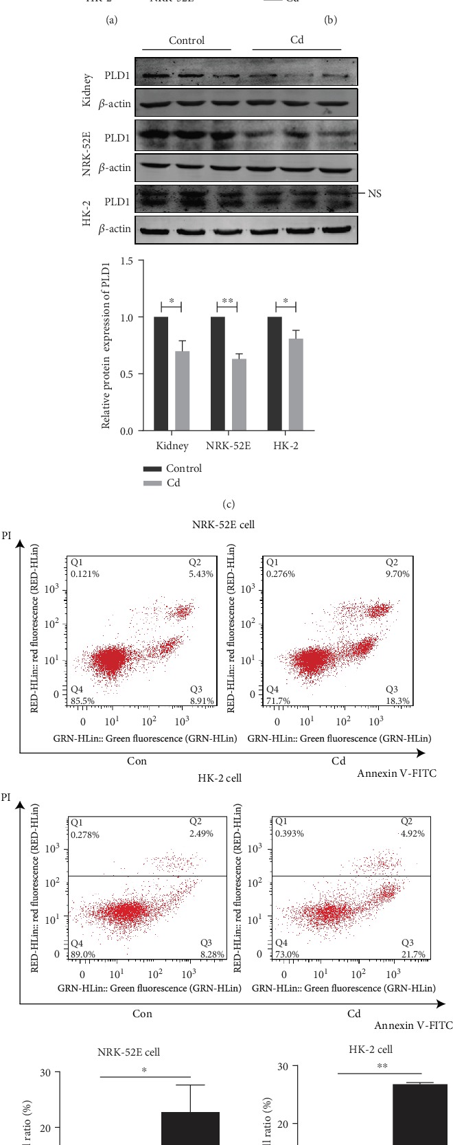
Figure 3.

PLD1 expression is inhibited significantly by Cd exposure in renal tubular cell lines. (a) The viability of NRK-52E and HK-2 cells after Cd exposure measured by CCK-8 assay. (b) The mRNA expression of PLD1 was decreased after Cd exposure. (c) The protein expression of PLD1 was decreased after Cd exposure. (d) Apoptosis ameliorated in renal tubular cells exposed to Cd performed by flow cytometry. Con: control group. Rats exposed to Cd at 0.6 mg/kg/d for 5 days per week for 12 weeks. NRK-52E cells exposed at 8 μM CdCl2 for 48 h, HK-2 cells exposed at 40 μM CdCl2 for 48 h. NS: nonspecific band. Data are mean ± SD. ∗p < 0.05 and ∗∗p < 0.01.
