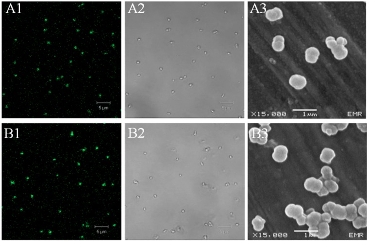
Figure 2.
Microscopic images of HSA-MPs (A1–3) and DOX-HSA-MPs (B1–3). (A1,A2) Confocal Laser Scanning Microscopy (CLSM) images of HSA-MPs and (B1, B2) CLSM images of DOX-HSA-MPs in fluorescence mode (excitation wavelength 488 nm, long pass emission filter 515 nm) and transmission mode, respectively; (A3,B3), SEM images of HSA-MPs and DOX-HSA-MPs, respectively.

