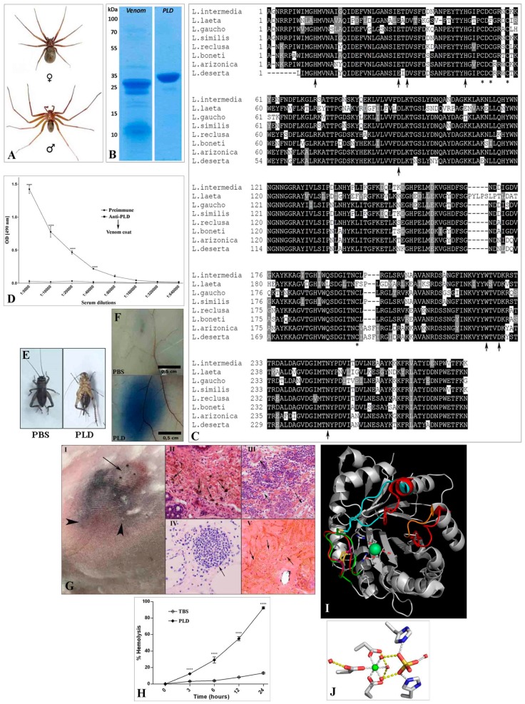
Figure 1.
Loxosceles phospholipases-D: general aspects and biological activities. (A) Sexual dimorphism between female and male adult L. intermedia specimens. (B) SDS-PAGE under reduced conditions of L. intermedia venom (10 µg) and phospholipase-D (PLD) LiRecDT1 (5 µg). (C) Multiple sequence alignment of representative phospholipases-D of L. intermedia (GenBank accession number ABA62021), L. laeta (AY093599), L. gaucho (JX866729), L. similis (AAX78234), L. reclusa (AY862486), L. boneti (AY559844), L. arizonica (AY699703) and L. deserta (C0JAU5). Sequences were aligned using the CLUSTAL X2 program [19]. Amino acid identities are shaded in black. Conservative substitutions are in gray, arrows point to amino acid residues involved in catalysis. The asterisks indicate cysteine residues. (D) Reactivity against L. intermedia crude venom using different dilutions of anti-LiRecDT1 serum (Anti-PLD) accessed by ELISA. The average ± standard errors are shown, with significance levels ****p ≤ 0.0001 comparing pre-immune with anti-PLD sera. (E) Representative images of crickets (n = 5) treated with Phosphate Buffered Saline (PBS) or LiRecDT1 (L. intermedia PLD, 4 μg) injected in the second segment of abdomen. (F) Increasing of vascular permeability of cutaneous blood vessels in mice triggered by LiRecDT1. (G) Dermonecrosis and histopathological changes following LiRecDT1 injection in rabbits’ tissue. (I) 5 µg of LiRecDT1 was injected subcutaneously in back skin. Arrow shows the site of injection and arrowheads show spreading of dermonecrotic lesion after 24 h. (II–V) Histopathological findings of rabbits’ skin 24 h following LiRecDT1 exposure. (II) Arrows show necrotic sites; (III) massive inflammatory response into the dermis and disorganization of collagen fibers pointed by arrows; (IV) massive inflammatory cell accumulation within dermal blood vessels (arrow); (V) hemorrhagic sites into the dermis are pointed by arrows. (H) Time-dependent direct hemolysis activity in rabbit erythrocytes treated with Phospholipase D of Loxosceles gaucho. (I) Cartoon representation of the structures of Brown spider venom PLDs: structural features highlighted in green, cyan and orange (catalytic, flexible, and variable loops, respectively) are from Loxosceles intermedia (PDB code: 3RLH) and in red are from Loxosceles laeta (PDB code: 1XX1). Amino acids participating in binding to Mg2+ (green sphere), catalysis and disulfide bridge formation are included in atom colors. (J) Mg2+ coordination (green sphere) by amino acid side chains and solvent molecules. Bound sulfate ion (yellow, red) is included. All procedures involving animals were carried out in accordance with “Brazilian Federal Laws”, following the Institutional Ethics Committee for Animal Studies Guidelines from Federal University of Paraná (Certificate n° 1112 of the Federal University of Paraná).

