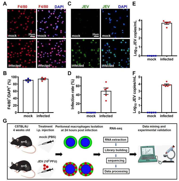
Figure 1.
Infection of the peritoneal macrophages and transcriptomic research design. Four-week-old female C57BL/6J mice were randomly divided into a mock-treated group (n = 5) and a Japanese encephalitis virus (JEV)-infected group (n = 5). At 24 hpi, mice were euthanized, and their sera, peritoneal macrophages, and peritoneal lavages were collected. (A) Immunofluorescent (IF) staining of F4/80 (red), a specific marker for macrophages. DAPI denotes the nucleus; scale bar = 20 μm. (B) Macrophage purity is shown as the proportion of F4/80+ cells. (C) IF staining of JEV antigens (green). DAPI denotes the nucleus; scale bar = 20 μm. (D) Infection rate of the peritoneal macrophages. (E) JEV RNA load in peritoneal lavages (each mouse was washed with 4 mL PBS). (F) JEV RNA load in mouse sera. (G) Schematic description of the transcriptomic research design. The quantitative results are shown as the mean ± SEM, and each dot denotes an independent mouse.

