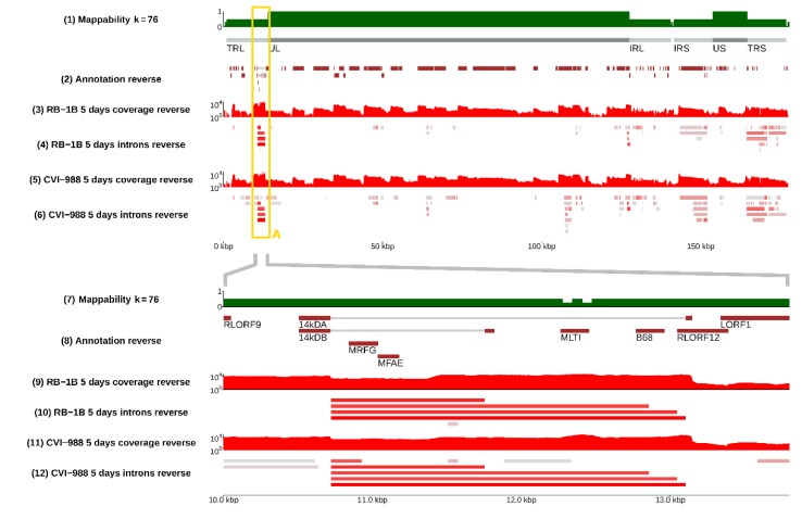
Figure 2.
The splicing landscape at the junction between terminal repeat regions. Top panel: negative strand of the whole MDV genome; bottom panel: region from 10 to 14 kb (TRL/UL junction). The bottom panel is a magnified version of the content of the yellow frame A present in the top panel and in Figure 1. Alternative spliceforms across the 14KD polypeptide (pp14) gene of MDV in strains RB-1B and CVI-988 are compared. From the RNA-sequencing signal, one can see four main alternative spliceforms in CEF cells infected by RB-1B, whereas five alternative spliceforms were identified in cells infected by CVI-988. As shown in track (8) (annotation), only two isoforms (14 kDA and 14 kDB) were present in the standard MDV annotations, as defined by Hong and Coussens [40]. The bottom panel of this figure can be reproduced in the online MDV genome browser by accessing https://mallorn.pirbright.ac.uk/browsers/MDV-annotation/Figure-2.html.

