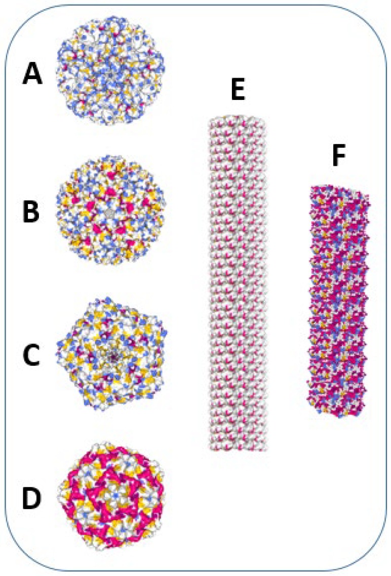
Figure 2.
Examples of icosahedral and helical plant viruses, used for vaccine development. Images were created using Protein Data Bank and NGL 3D viewer [27]. α-helices are shown in red, β-sheets–in yellow. (A) Cucumber mosaic virus (CMV) structure (T = 3 symmetry, diameter = 28 nm). Image of 5OW6 [28]. (B) Cowpea chlorotic mottle virus (CCMV) structure (T = 3 symmetry, diameter = 29 nm). Image of 1CWP [29]. (C) Cowpea mosaic virus (CPMV) structure (T = 3 symmetry, diameter = 28 nm). Image of 1NY7 [30]. (D) Sesbania mosaic virus (SeMV) structure (T = 3 symmetry, diameter = 28 nm). Image of 1X33 [31]. (E) Tobacco mosaic virus (TMV) structure (cryo-EM reconstruction of a TMV fragment; particle length = 300 nm, diameter = 18 nm). Image of 3J06 [32]. (F) Bamboo mosaic virus (BaMV) structure (cryo-EM reconstruction of a BaMV fragment; particle length = 490 nm, diameter = 15 nm). Image of 5A2T [33].

